Sloane Kelly ’22 has taken college science classes before, the kind where lectures and labs are held separately and on different days. This fall, however, the health promotion major at University of Lynchburg is taking a new pilot course in human anatomy and physiology in which the lecture and lab are combined.
“I absolutely love it,” Kelly said. “Especially with a subject such as anatomy and physiology, it’s extremely helpful to learn fundamental information in lecture and then immediately apply it in a lab, as Dr. Williamson often does.
“I have been in science classes before, where it was a separate three-hour lab, once a week. While I learned the material in lecture and lab, the course material didn’t seem as cohesive between lecture and lab as it does with the integrated structure.”
The class, which meets for two hours on Mondays, Wednesdays, and Fridays, is taught by Dr. Wendy Williamson, who is a physical therapist and an assistant professor in the School of Sciences. It’s a requirement for students in a number of majors, including nursing, exercise physiology, and other health science-related programs.
“Last year, I took chemistry and chemistry lab, separated, and it was challenging,” health promotions major Emma Drake ’22 said. “I do like the integrated [class and lab] more because while in lecture we’re learning about it and then right after we learn it we are getting hands-on experience in the lab.
“It’s nice, being able to learn from lecture and lab, back to back. It does get tiring, but Dr. Williamson is good on giving us breaks and keeping us engaged.”

Williamson said she hopes all of her students are enjoying the new format. “I aim to make it student centered, with the ability to move from lecture-based content to kinesthetic opportunities within the same class period in the same space,” she said. “There is certainly a level of autonomy and responsibility I expect from my students.
“I try to have a good mix of didactic material, clinically relevant examples, and hands-on experiences, and I think the students will benefit from not having to wait a week until the next lab, as in the other model, to apply what they’ve learned. The coordination of lecture and lab materials is very complementary and this will benefit students as well.”
The class meets in a newly renovated space at Hobbs-Sigler Hall, the University’s science building. This past summer, several areas of Hobbs-Sigler were refurbished, including the anatomy and physiology lab. It’s now an active learning classroom, with furniture that can be easily rearranged, flat-screen monitors, and dry-erase boards.
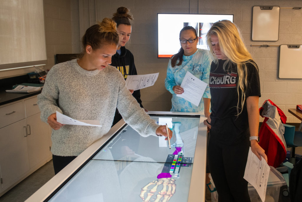
It also includes an Anatomage Table, which has been billed as “the world’s first virtual dissection table.” It looks like a giant tablet computer, albeit one that’s loaded with a variety of 3D medical images.

The Table supplements the University’s undergraduate cadaver lab, one of two cadaver labs on campus, and enables students to do the kind of research that once required an actual cadaver, something that’s not an unlimited resource.
“A lot of medical schools are starting to go this way,” Dr. William Lokar, dean of the Lynchburg College of Arts and Sciences, said. “I don’t even know how many graduate health programs have these. Wendy Williamson brought it to my attention a while back and we started talking about it and it made sense for a lot of reasons.”
Kelly said being able to learn about the human body in 3D, with the Anatomage Table, instead in 2D, from a textbook, has been better. “While a book can give you all the details you would ever need to know, seeing a 3D image puts anatomy into perspective and makes it a reality,” she said. “Humans are not 2D drawings on a page, therefore, it’s helpful to learn anatomy in a 3D manner.”
Drake also prefers the Anatomage Table to a textbook, saying it’s “a lot easier” and enables her to “explore the human body” without having to use a cadaver. “I love being able to work on the Table and being so hands on provides me with a great learning experience,” she said.
Williamson said having the Anatomage Table has been a positive experience for her students. “They catch on very quickly and manipulating the images is very intuitive,” she said. “I think they enjoy using technology, seeing the structures in different ways, and the ability to explore.
“The most interesting moment has been when we pulled up the specific pathologies of the cadavers to see what medical issues they had going on at the time of death. For example, one had lung pathology from smoking. Another had gastrointestinal cancer with a fistula and metastases to the spine with a compression fracture.
“We tied those real examples to what we were learning in class about the vertebral column. It also helped my students realize that real people were used to create the 3D images.”
Drake, who plans to pursue a master’s degree with the goal of becoming a health care administrator, said the integrated lecture/lab and Anatomage Table are giving her a head start. “Taking this class and being able to work on the Table is providing me with an amazing learning experience,” she said.
“I am able to go inside the body and learn about that person with a touch of a few buttons. I think working with the Table is great because our world is turning into a technology-based world, and learning how to work with this piece of equipment so early is going to be very helpful.”

