Billed as “the world’s first virtual dissection table,” the Anatomage Table looks like a giant tablet computer, albeit one that’s loaded with a variety of cadavers and other anatomical and medical images.
“It includes a variety of cadavers, histology images, pathophysiology, dynamic anatomical functions including blood flow through the body and respiration, and endoscopic procedures,” Dr. Wendy Williamson, who teaches human anatomy and physiology at Lynchburg, said. “Students will even have the ability to input their own radiologic images to view on the Table.”
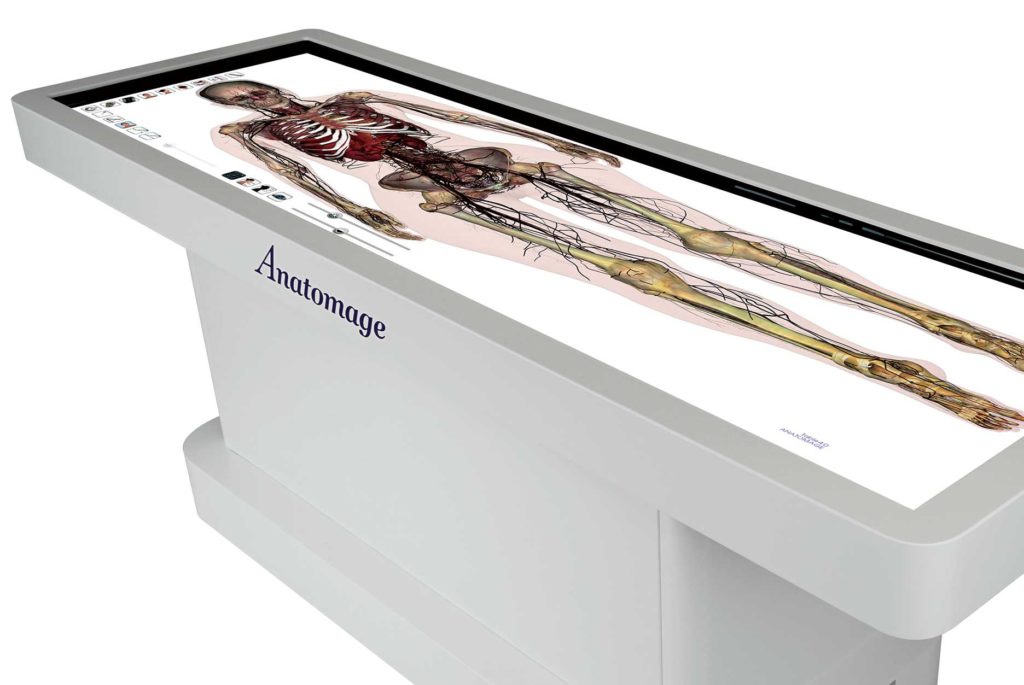
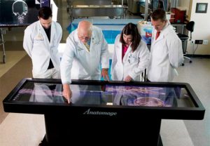
Images on the Table can be manipulated, too, giving students the opportunity to study the human body and its processes layer by layer. “The Anatomage table has an 81-by-28-inch touch screen, encouraging intuitive manipulation of images, as well as multi-plane dissection and viewing of virtual cadavers,” Dr. Williamson said.
“The study of anatomy and physiology is full of complex, dimensional relationships and the Anatomage Table allows for systemic investigation and discovery of these associations.”
The Table will be used in Lynchburg’s undergraduate human anatomy and physiology classes, which are required for students in a number of majors, including nursing, exercise physiology, biology, biomedical science, and other health science-related programs. About 140 to 150 students take anatomy and physiology each year.
The Table will supplement the University’s undergraduate cadaver lab, one of two cadaver labs on campus. It will enable students to do the kind of research that once required an actual cadaver, something that’s not an unlimited resource.
“A lot of medical schools are starting to go this way,” Dr. William Lokar, Dean of the Lynchburg College of Arts and Sciences, said of the Table. “I don’t even know how many graduate health programs have these. Wendy Williamson brought it to my attention a while back and we started talking about it and it made sense for a lot of reasons.”
The Table also will be used in the Peer-Assisted Supplemental Study program, a widely used resource at Lynchburg. PASS leaders attend class lectures in traditionally difficult classes and then tutor other students, one-on-one or in group settings.
“It will be an excellent opportunity for our PASS sessions,” Dr. Lokar said. “Students will have access to these virtual cadavers at hours when supervised work with human cadavers isn’t possible. It’s a new pedagogical tool that we feel will improve student learning.”
Dr. Williamson, who will attend the Anatomage Table Users Group Meeting in California next month, called the Table a “tremendous asset” for students. “It’s very visually stimulating and would allow our students to increase time-on-task in a novel and innovative way,” she said.
“Learning styles vary greatly within our diverse student population, and students report improved understanding of anatomy and physiology when a variety of learning opportunities are provided. In addition, college-level students have been shown to benefit from problem-based learning, using patient scenarios for the application of didactic anatomy and physiology content.”
Faculty at Lynchburg also are looking forward to using the Table. “Instructors at Lynchburg are highly motivated to use interactive and engaging technology in the classroom to enhance our students’ experiences,” Dr. Williamson said. “The Table would create opportunities for increased variety of instructional methods shown to be effective in learning.
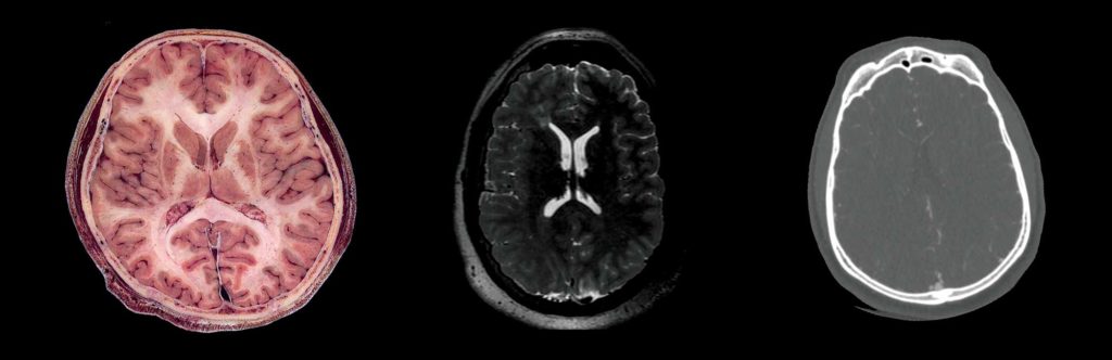
“Images from the Table can be imported into lecture material to ensure consistent content. Pathology images available on the Table allow faculty to fully incorporate patient scenarios into instruction and testing.
“The high-resolution imaging available on the Table allows for extremely detailed visualization and can be implemented in a variety of student-faculty research collaborations.”
Dr. Williamson envisions a community outreach component, too.
“In addition to the benefits to our students and faculty, I’m enthused about the opportunity for community outreach,” she said. “I would like to invite practicing health care professionals, graduate students, and local high school anatomy and physiology classes to take advantage of educational opportunities using the Table.”
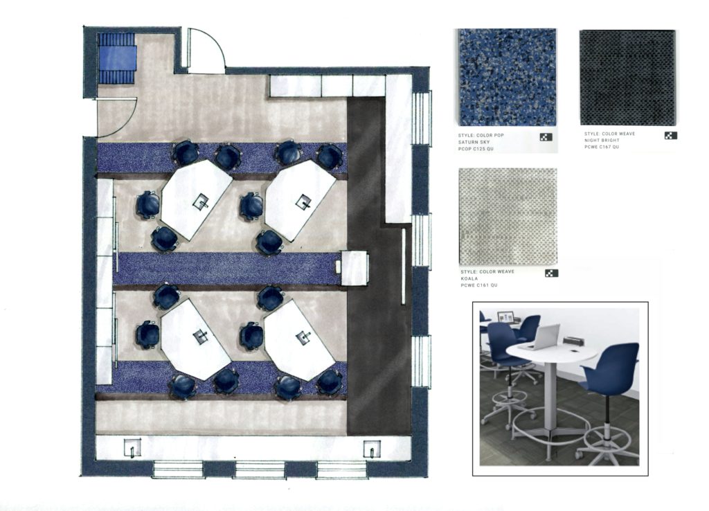
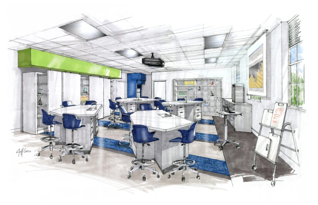
The Anatomage Table will be housed in a newly renovated space at Hobbs-Sigler Hall. Several areas of the science building are being renovated this summer, including the anatomy and physiology lab, where the Anatomage Table will be located.
When work is completed later this summer, the lab also will be an active-learning classroom with furniture that can be easily rearranged, flat-screen monitors, and dry-erase boards.
“The renovation to our Hobbs classroom includes not only the addition of the Table, but also an exciting transition to an active-learning classroom,” Dr. Williamson said. “This will facilitate collaborative work, hands-on interactions, and peer-to-peer instruction that are vital to maintaining student engagement and success.”
Other improvements being made to Hobbs-Sigler include renovating the microbiology lab, transforming two classrooms into a new conference room, and making a small research lab that was previously accessible only through a faculty member’s office more easily accessible. Work is expected to be completed by the beginning of the Fall semester.
“It will be a more modern space that will allow us to work with our students,” Dr. Lokar said.
Also this fall, Dr. Williamson will begin a pilot course in which the anatomy and physiology lectures and labs are integrated into one course. Instead of attending three one-hour lectures per week and one three-hour lab, students in the pilot course will meet two hours at a time, three days a week, for sessions that involve lectures and lab work.
“I think students will benefit greatly from both the new technology and innovative changes in teaching the content,” Dr. Williamson said.